
(Part 1)The 3rd Huanhu Skull Base Surgery Conference Concludes Successfully
The 3rd Huanhu Skull Base Surgery Conference 2026
On the final day of the 3rd Huanhu Skull Base Surgery Conference 2026, the scientific program focused on practical surgical strategies for managing complex skull base lesions, with in-depth discussions on the meticulous prevention and management of surgical complications.
1. the launch ceremony of the Huanhu Skull Base Academy
The launch ceremony was presided over by Professor Wang Hongguang. Professors Juan Fernández -Miranda, Eric W. Wang, Pablo Augusto Rubino, Jeffrey Sorenson, Georgios Zenonos, Martin Jurlina, Tong Xiaoguang, Liu Gang, and Wang Hongguang jointly participated in the Launch Ceremony of the Huanhu Skull Base Academy, witnessing this significant milestone together.
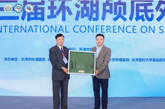
Professor Su Ning from the Department of Neurosurgery at the People's Hospital of Inner Mongolia Autonomous Region, on behalf of the widow of the late Professor Takanori Fukushima, presented Professor Tong Xiaoguang of Tianjin Huanhu Hospital with a complete set of microsurgical instruments developed personally by Professor Fukushima. This meaningful gesture not only honored Professor Fukushima's profound influence on microneurosurgery but also conveyed recognition of Professor Tong Xiaoguang's distinguished standing and contributions in the field of neurosurgery.
2. Scientific Presentations
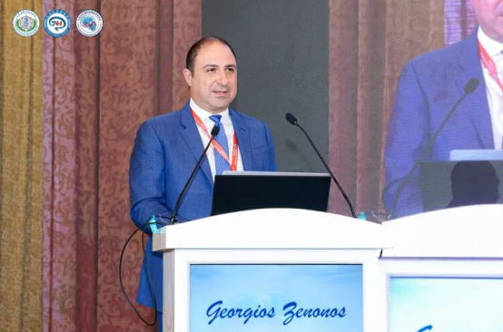
Professor Georgios Zenonos delivered a presentation entitled “The 360 Approach for the Treatment of Skull Base Pathology,” exploring comprehensive, multidirectional strategies for managing complex skull base lesions. He introduced cutting-edge techniques in skull base surgery, particularly the expanded boundaries of the endoscopic endonasal approach, including access to the lateral cavernous sinus and lateral mobilization of the internal carotid artery. He also discussed the combined extreme medial transcondylar, transjugular tubercle, and transmastoid approaches for recurrent chordomas; the far-lateral approach for ruptured posterior inferior cerebellar artery aneurysms; and advanced cerebrovascular bypass and reconstruction techniques. Professor Zenonos emphasized that complex skull base lesions often require a combination of transcranial and endoscopic endonasal approaches. He highlighted the complementary roles of ventral and lateral skull base surgery and cerebrovascular surgery, advocating their coordinated integration to achieve optimal surgical outcomes.
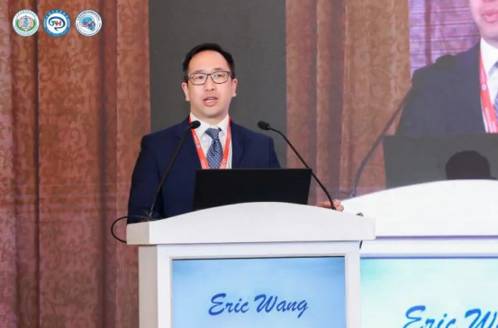
Professor Eric W. Wang delivered a presentation entitled “Complications of Endoscopic Skull Base Surgery,” addressing the challenges associated with complications of endoscopic procedures, including postoperative saddle-nose deformity, olfactory dysfunction, and cerebrospinal fluid (CSF) leakage. He discussed techniques for preserving the olfactory mucosa during nasoseptal flap harvesting, key considerations in the repair of severe CSF leaks, and strategies for preventing abducens nerve palsy. His presentation provided valuable practical insights into minimizing surgical complications and enhancing patient safety in endoscopic skull base surgery.
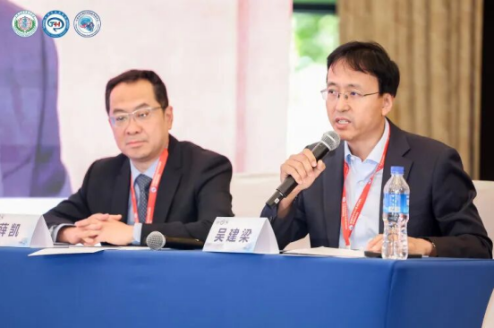
The scientific presentation session was co-chaired by Professor Wu Jianliang and Professor Xue Kai.
3. Chordoma Session
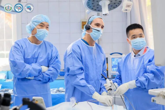
Professor Juan Fernández -Miranda delivered a detailed endoscopic anatomical demonstration focusing on the clivus and surrounding structures of the jugular foramen. He began by creating a U-shaped incision in the posterior pharyngeal wall, detaching the attachments of the longus capitis muscles, and reflecting the posterior pharyngeal fascial flap inferiorly to expose the lower clivus. He then demonstrated lateral exposure of the hypoglossal canal, occipital condyle, foramen magnum, and anterior arch of C1, highlighting key anatomical landmarks and surgical precautions.
He further illustrated a stepwise surgical technique, beginning with bone removal along the anterior margin of the foramen magnum at the midline, followed by lateral extension to expose the hypoglossal canal. Subsequent drilling of the jugular tubercle and opening of the dura mater allowed visualization of critical neurovascular structures within the cerebellopontine angle. The demonstration highlighted the unique advantages of the endoscopic endonasal approach in providing a medial perspective of the jugular foramen and cerebellopontine angle regions.
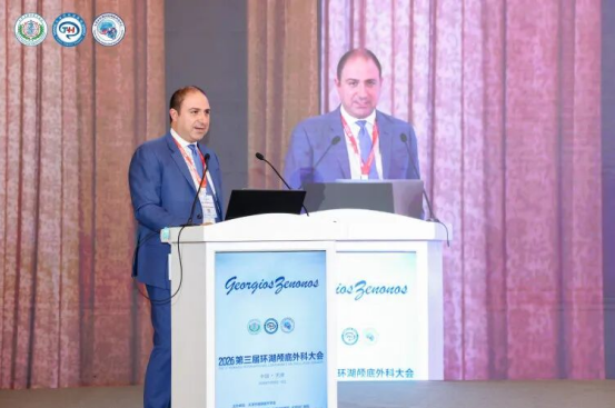
Professor Georgios Zenonos delivered a presentation entitled “Comprehensive Management of Clival Chordoma,” sharing the extensive experience of the University of Pittsburgh Medical Center (UPMC) in the multidisciplinary management of clival chordomas. His presentation covered innovations in surgical approaches, skull base reconstruction strategies, molecular pathological analysis and prognostic assessment, as well as clinical cases involving pharmacological treatment. He emphasized that the management of chordomas extends far beyond surgical intervention alone and requires close collaboration among multiple specialties, including neurosurgery, otolaryngology, medical oncology, radiation oncology, and pathology, to develop the most appropriate individualized treatment strategy for each patient.
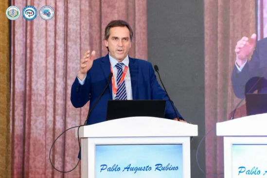
Professor Pablo Augusto Rubino presented a detailed layer-by-layer anatomical dissection of the far-lateral approach, accompanied by anatomical demonstrations and operative case presentations illustrating the surgical management of neoplastic and vascular lesions involving different regions of the brainstem and jugular foramen. He emphasized that the far-lateral approach provides an optimal surgical corridor for accessing and treating vascular and neoplastic lesions around the foramen magnum. He concluded that a thorough understanding of the intricate anatomy of this region is essential for the successful surgical management of complex lesions involving these critical structures.
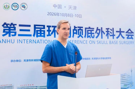
Professor Juan Fernández -Miranda concluded that the endoscopic approach provides a ventral surgical corridor to the skull base, and that favorable surgical outcomes can be achieved by selecting the most appropriate approach according to tumor location and histopathological characteristics.
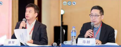
The scientific presentation session was co-chaired by Professor Kang Jun and Professor Ma Yue.