
(Part 2)The 3rd Huanhu Skull Base Surgery Conference Concludes Successfully
The 3rd Huanhu Skull Base Surgery Conference 2026
4. Jugular Foramen Tumor Session
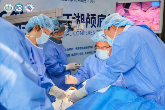
Professor Feng Guodong conducted an anatomical demonstration of the Fisch Type A Infratemporal Fossa Approach, the fundamental technique within the Fisch classification of infratemporal fossa approaches, primarily indicated for complex lesions involving the jugular foramen region. He outlined the surgical incision and essential operative steps, emphasizing that this approach is particularly suitable for jugular foramen lesions encasing the internal carotid artery.
He stressed that surgical anatomical dissection should be guided by practical operative scenarios rather than aimed merely at exposing anatomical structures. He further explained that subtotal petrosectomy is an independent surgical procedure for managing conditions such as cerebrospinal fluid (CSF) leakage and severe infections, while also constituting the initial stage of the infratemporal fossa approach. Exposure of approximately 270 degrees of the facial nerve canal is a critical prerequisite for anterior transposition of the facial nerve. He also highlighted the importance of tailoring the extent and site of facial nerve transposition according to the location of the lesion.
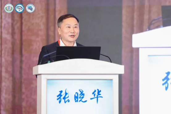
Professor Zhang Xiaohua presented a surgical strategy for achieving 360-degree exposure of the jugular foramen region, beginning with a representative case of jugular bulb meningioma. He demonstrated how the Fisch infratemporal fossa approach, combined with the far-lateral approach, enables circumferential exposure of the jugular vein and its surrounding structures. The Fisch approach provides access to the superior, anterior, and inferior aspects of the jugular bulb, whereas the far-lateral approach facilitates exposure of the posteroinferior region.
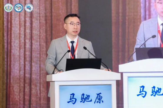
Professor Ma Chiyuan delivered a presentation entitled “Endoscopic Resection of Skull Base Lesions via Natural Ventral Corridors: Anatomical Considerations and Surgical Techniques.” He began by defining the relevant anatomical subdivisions of the skull base accessible through endoscopic endonasal, transoral, and transorbital approaches, enabling surgeons to localize lesions accurately under endoscopic visualization and select the most appropriate surgical corridors. Drawing on representative clinical cases, he demonstrated the key operative steps and technical nuances of endoscopic endonasal, transoral, and transorbital approaches for resecting lesions in different skull base regions. He further proposed that the integration of multiple endoscopic surgical corridors represents an important direction for the next stage of standardized skull base surgery.
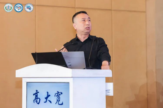
Professor Gao Dakuan delivered an engaging presentation demonstrating endoscopic endonasal techniques for internal carotid artery and Eustachian tube mobilization and their clinical applications. He emphasized that the ultimate goal of skull base surgery is to achieve adequate tumor exposure and maximal safe resection while preserving the function of critical vascular and neural structures to the greatest extent possible. He further highlighted the ability of endoscopic techniques to provide precise surgical exposure of the anteroinferomedial aspect of the jugular foramen region.
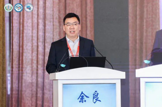
Professor Yu Liang provided a comprehensive overview of the extended middle cranial fossa approach for the resection of petroclival and brainstem lesions. He offered an in-depth analysis of the approach from three perspectives: its historical development, anatomical foundations, and key surgical techniques, illustrating his presentation with several representative clinical cases. In addition, Professor Yu shared historical photographs of himself and his colleagues attending training courses led by Professor Tong Xiaoguang, expressing his profound respect for the rich legacy of microneurosurgery at Tianjin Huanhu Hospital and his commitment to carrying forward its distinguished surgical traditions.
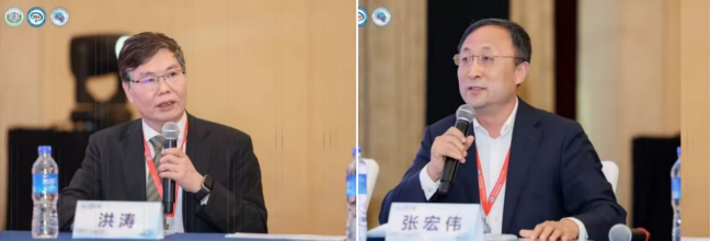
The scientific presentation session was co-chaired by Professors Hong Tao, Zhang Hongwei, Yao Yong, and Ma Yue.
Anatomical Summary
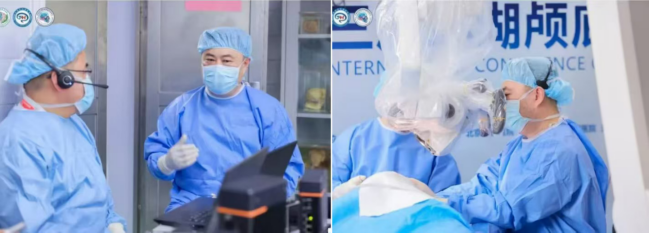
Professor Tong Xiaoguang provided a detailed anatomical demonstration of the jugular foramen region using the far-lateral approach. He systematically compared the neurosurgical far-lateral approach with the otolaryngological Fisch Type A infratemporal fossa approach, highlighting their respective characteristics and differences in surgical exposure of jugular foramen lesions. He further explored strategies for selecting the optimal surgical approach according to the specific anatomical and pathological characteristics of individual lesions, as well as the clinical application of combined approaches for the effective management of complex jugular foramen pathology.
5. Huanhu Skull Base Academy Pituitary Tumor Masterclass
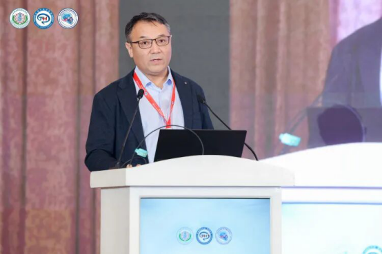
Professor Serik Duysenbi from Kazakhstan presented his experience in managing skull base tumors encasing major blood vessels, demonstrating how vascular reconstruction performed before tumor resection can achieve favorable clinical outcomes. He also emphasized his commitment to advancing skull base neurosurgery across Central Asia, with Kazakhstan serving as a regional hub. Through close collaboration with Professor Tong Xiaoguang and his team, he outlined plans to promote the sustainable development of the specialty by establishing systematic development strategies and placing particular emphasis on the training of young neurosurgeons.
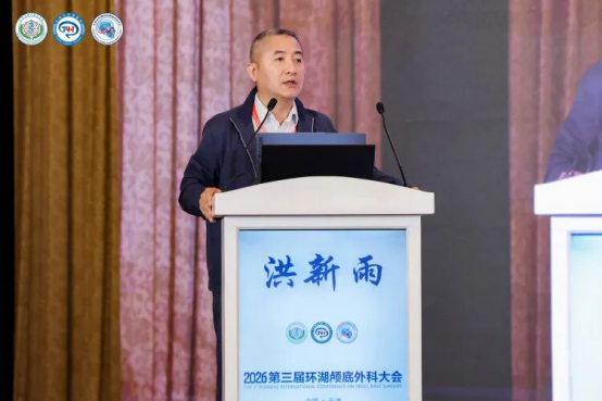
Professor Hong Xinyu shared his clinical experience in the endoscopic surgical treatment and management of craniopharyngiomas, highlighting the role of intraoperative visual evoked potential monitoring in reducing the incidence of postoperative visual deterioration. Drawing on representative clinical cases, he emphasized that craniopharyngioma resection should follow the principles of “debulking first, identifying tumor boundaries, in situ dissection, and avoiding traction.” For tumors adherent to the optic nerves, he recommended initial blunt dissection, followed by sharp dissection when necessary, while maintaining meticulous protection of the hypothalamus and critical neurovascular structures throughout the procedure.
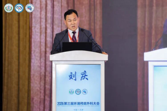
Professor Liu Qing delivered a presentation entitled “Reflections and Clinical Practice on Surgical Treatment Selection for Craniopharyngiomas in the Endoscopic Era.” He emphasized the importance of critically evaluating the concept of adopting the endoscopic endonasal approach as the first-line surgical option for craniopharyngiomas, while continuing to explore and refine transcranial surgical approaches. He advocated radical tumor resection with preservation of hypothalamic–pituitary function and highlighted the implementation of individualized surgical strategies based on his pioneering ten-subtype classification system for craniopharyngiomas.
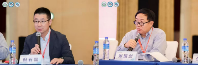
The scientific presentation session was co-chaired by Professor Ni Shilei and Professor Zhang Ruijian.
6. Scientific Presentations
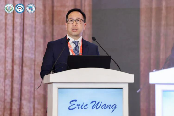
Professor Eric W. Wang delivered a presentation on transpterygoid, transmaxillary, and contralateral transmaxillary approaches to the pterygopalatine fossa and petrous apex. He highlighted that the transpterygoid approach is suitable for managing lesions involving the pterygopalatine fossa, petrous apex, and nasopharynx, serving as an important complement to approaches targeting the clivus. For lesions of the petrous apex, he emphasized that the contralateral transmaxillary (CTM) approach may offer a more favorable surgical corridor.
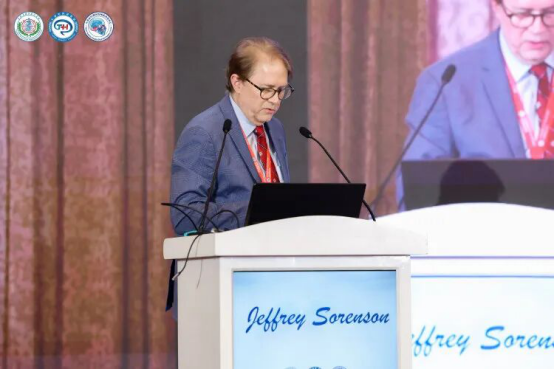
Professor Jeffrey Sorenson presented a step-by-step demonstration of the postauricular transtemporal approach to the jugular foramen, using anatomical animations, photographs, and representative clinical cases. He detailed each stage of the surgical approach, including muscle dissection, vascular exposure and management, and bone removal.
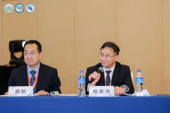
The scientific presentation session was co-chaired by Professor Lou Meiqing and Professor Xue Kai.