
(Part 3)The 3rd Huanhu Skull Base Surgery Conference Concludes Successfully
The 3rd Huanhu Skull Base Surgery Conference 2026
7. Endoscopic Anatomy
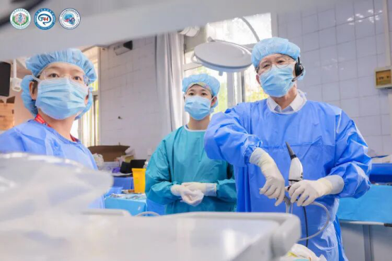
Professor Jiang Weihong conducted an endoscopic anatomical demonstration of the Eustachian tube region. Through endoscopic drilling of the posterior wall of the maxillary sinus and the pterygoid process, he exposed the fascia covering the pterygoid venous plexus, the maxillary division of the trigeminal nerve (V2), and the levator veli palatini muscle, thereby identifying the parapharyngeal space and Eustachian tube. He subsequently demonstrated resection of the Eustachian tube and the fascial structures surrounding the internal carotid artery at the foramen lacerum, allowing mobilization of the internal carotid artery.
8. Nasopharyngeal Carcinoma Session
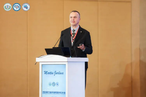
Professor Martin Jurlina delivered a presentation on “Surgical Techniques and Challenges in the Management of Malignant Tumors of the Parapharyngeal Space.” He emphasized that meticulous preoperative surgical planning is essential and that surgical strategies should not be improvised intraoperatively. He explained that transmandibular and lateral orbitozygomatic approaches can facilitate the resection of malignant parapharyngeal space tumors with favorable clinical outcomes. In contrast, he stressed that achieving negative surgical margins through an endoscopic transmaxillary approach is not feasible for large malignant tumors involving the parapharyngeal space. At the conclusion of his presentation, he once again extended an invitation to all attendees to participate in the upcoming European Skull Base Congress in London.
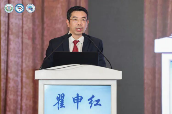
Professor Qu Shenhong presented the clinical characteristics of recurrent nasopharyngeal carcinoma in high-incidence regions from a surgeon’s perspective. He emphasized that extensive skull base necrosis requires intraoperative adjustment and expansion of the resection margins according to the extent of necrotic tissue involvement. He also highlighted that pathological changes in the mechanical properties of the affected internal carotid artery wall may increase the risk of intraoperative hemorrhage. Furthermore, he noted that cerebrospinal fluid (CSF) leakage following radiotherapy remains a significant surgical challenge and identified the vascularized temporalis muscle flap as a valuable option for skull base reconstruction.
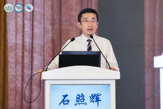
Professor Shi Zhaohui presented his clinical experience and evolving treatment strategies for recurrent nasopharyngeal carcinoma complicated by tissue necrosis. He outlined three fundamental principles for the surgical management of necrotic lesions: infection control, tissue repair and reconstruction, and prevention of complications. He emphasized that recurrent nasopharyngeal carcinoma accompanied by extensive necrosis poses particularly complex therapeutic challenges, requiring careful determination of the sequence of surgical and medical treatments according to the severity of necrosis. He further highlighted the importance of tailoring reconstructive strategies to the extent of tissue involvement, identifying well-vascularized pedicled muscle flaps and free tissue flaps as effective options for repairing complex defects.
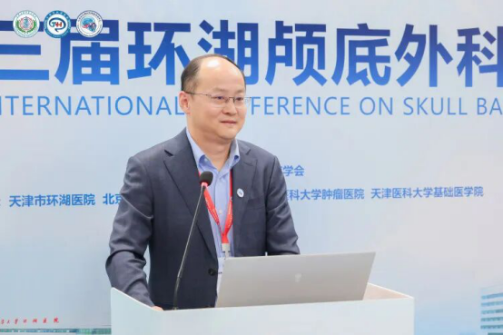
Professor Zhai Xiang presented the management strategies developed at Tianjin Huanhu Hospital for vascular injuries resulting from skull base necrosis following radiotherapy for nasopharyngeal carcinoma. He emphasized that the management of major skull base vessels, particularly the internal carotid artery, is a critical consideration in skull base surgery. Regarding internal carotid artery hemorrhage, he explained that his team prioritizes vascular bypass and reconstruction when occlusion of the affected artery is not feasible. Drawing on a representative clinical case of internal carotid artery hemorrhage caused by radiation-induced skull base necrosis following treatment for nasopharyngeal carcinoma, he highlighted the importance of multidisciplinary collaboration and sound clinical decision-making in managing these complex and potentially life-threatening conditions.
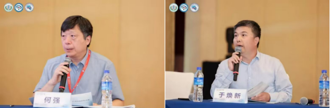
The scientific presentation session was co-chaired by Professors Zhou Bing, He Qiang, Wang Zhaoyan, and Yu Huanxin.
9. Parapharyngeal Space and Head and Neck Surgery Session
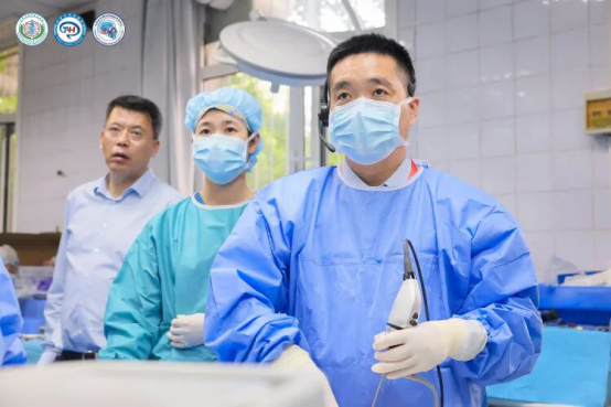
Professor Sun Xicai conducted a step-by-step anatomical demonstration of the endoscopic transoral approach to parapharyngeal space lesions. Beginning with the surgical incision, he demonstrated dissection through the tissue planes between the superior pharyngeal constrictor muscle and the temporalis muscle to access the prestyloid compartment. Following division of the styloid diaphragm, he exposed key anatomical structures, including the stylopharyngeus muscle, glossopharyngeal nerve, carotid sheath, longus capitis muscle, hypoglossal nerve, and vagus nerve. The demonstration provided a comprehensive overview of the surgical anatomy and exposure of the parapharyngeal space through the endoscopic transoral approach.
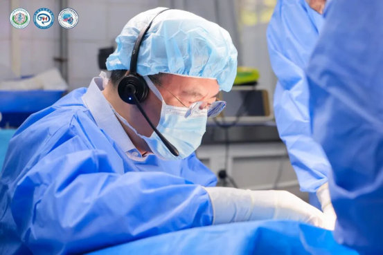
Drawing on his extensive clinical and anatomical experience, Professor Wang Xudong demonstrated the surgical management of parapharyngeal space lesions via the lateral transcervical approach. He emphasized that accurate identification, meticulous dissection, and preservation of the facial nerve are essential prerequisites for safely accessing the parapharyngeal space through this approach.
10. Closing Ceremony
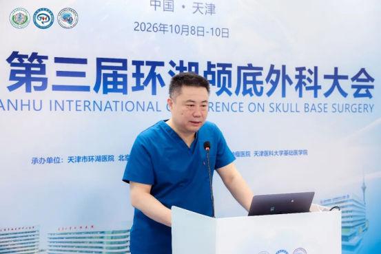
Professor Tong Xiaoguang delivered the concluding remarks. He highlighted that, over the years, the Skull Base Surgery Center at Tianjin Huanhu Hospital has consistently upheld a distinctive conference format integrating real-world surgical case presentations with live anatomical demonstrations. By inviting renowned experts from China and abroad, the conference has provided a comprehensive platform for showcasing authentic skull base surgical techniques and sharing practical clinical expertise.
Reflecting on the two-and-a-half-day conference, Professor Tong praised the rich and engaging scientific program and expressed his appreciation for the dedication and enthusiastic participation of all attendees. He extended his sincere gratitude to everyone for attending the conference and to the distinguished speakers for generously sharing their knowledge, experience, and surgical expertise.
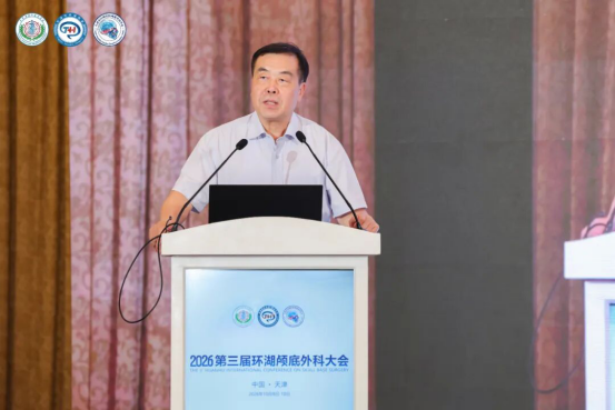
Professor Liu Gang delivered the closing address, expressing his heartfelt gratitude to the distinguished experts from China and abroad for their outstanding academic presentations and hands-on demonstrations of anatomical approaches. He also extended his sincere appreciation to the Department of Anatomy at Tianjin Medical University for providing the anatomical specimens essential to the success of the conference.
Professor Liu announced that the 4th Huanhu Skull Base Surgery Conference would be held in Tianjin in 2027 and warmly invited colleagues from around the world to reunite in the city for further academic exchange, collaborative learning, and discussions on advances in skull base surgery.
Finally, Professor Liu Gang formally declared: “The 3rd Huanhu Skull Base Surgery Conference is officially concluded!”