
(Part 3)【Grand Opening】Integrating Cutting-Edge Technologies, Exploring New Frontiers in Skull Base Surgery — Highlights Continue at the 3rd Huanhu Skull Base Surgery Conference
04 Petroclival Region Session
Professor Juan Fernandez-Miranda and Professor Sun Xicai once again joined forces to deliver a themed presentation and hands-on anatomical demonstration entitled “Endoscopic Anatomy of the Petrous Apex Region.” Through detailed demonstrations of the intricate anatomy of the petrous apex under endoscopic visualization, they highlighted the spatial relationships between neural and vascular structures in the petroclival region and delineated the anatomical limits of surgical exposure achievable through the endoscopic endonasal approach. Their presentation provided an essential anatomical foundation for the endoscopic surgical management of complex petroclival tumors.
Professor Zhang Hongwei presented a combined endoscopic and microsurgical approach, tailoring surgical strategies according to the extent of tumor invasion. For giant pituitary adenomas extending into the cavernous sinus and petroclival region, he leveraged the superior visualization of deep anatomical structures afforded by endoscopy to optimize surgical approach selection.
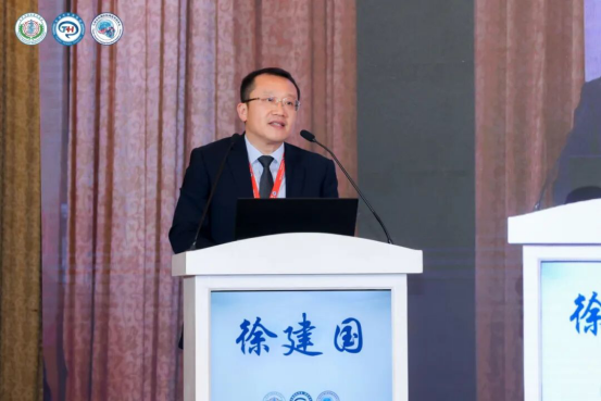
Professor Xu Jianguo highlighted the complementary roles of endoscopy and microscopy in the surgical management of petroclival lesions. By utilizing endoscopy to visualize deep anatomical regions and blind spots inaccessible under the microscope, while employing microsurgery to manage more superficial structures, this combined approach aims to improve gross total resection rates for petroclival lesions and reduce the risk of surgery-related neurological injury.
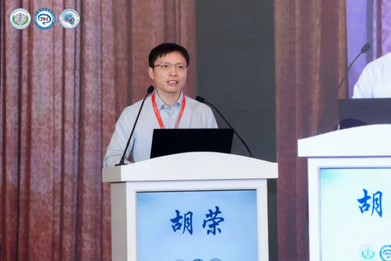
Professor Hu Rong discussed the endoscopic endonasal surgical management of various skull base tumors invading the cavernous sinus. He emphasized the importance of assessing the degree of tumor invasion and carefully defining the limits of endoscopic resection to achieve maximal safe tumor removal while substantially reducing the risk of postoperative complications.
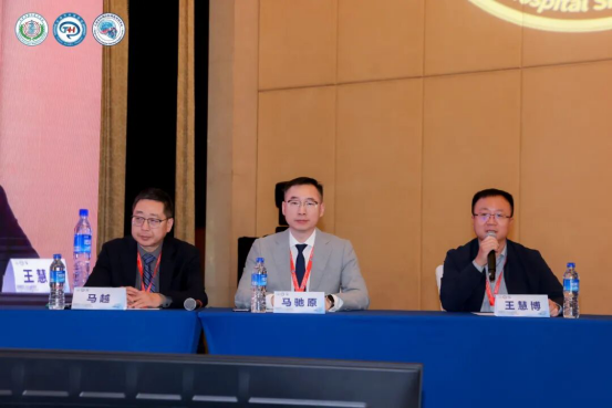
The scientific presentation session was co-chaired by Professors Ma Chiyuan, Wang Huibo, and Ma Yue.
05 Vestibular Schwannoma Session
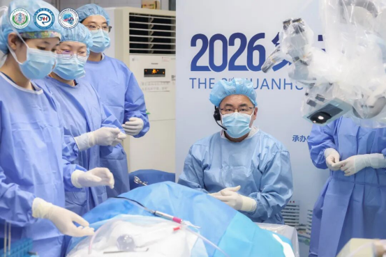
Professor Feng Guodong opened the session with a presentation entitled “Anatomy of the Internal Auditory Canal via the Middle Cranial Fossa Approach: An Otolaryngological Perspective.” Drawing on the interdisciplinary integration of otology and skull base surgery, he provided a detailed overview of the key anatomical landmarks of the internal auditory canal via the middle cranial fossa approach, highlighting the anatomical courses of the facial and vestibulocochlear nerves. His presentation provided an essential anatomical foundation for endoscope-assisted vestibular schwannoma surgery and hearing preservation.
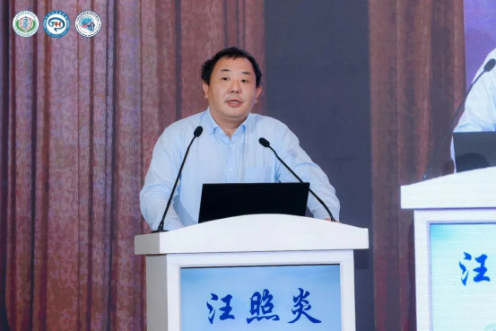
Professor Wang Zhaoyan presented the translabyrinthine approach to vestibular schwannoma surgery, highlighting its ability to provide extensive exposure of the internal auditory canal. He discussed the indications, advantages, and limitations of this approach, emphasizing the importance of protecting the sigmoid sinus and jugular bulb during surgery. He also compared the technical characteristics of the translabyrinthine approach with those of conventional neurosurgical approaches and shared practical techniques for tumor debulking using hydrodissection to facilitate nerve preservation. In addition, he introduced various techniques for nerve reconstruction following facial nerve injury, with particular emphasis on maximizing the preservation and restoration of facial and auditory nerve function.
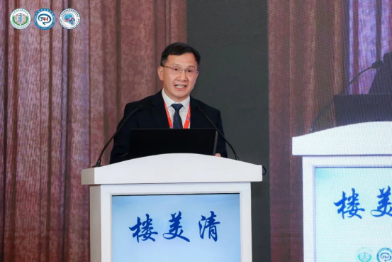
Professor Lou Meiqing introduced the concept of establishing a specialized vestibular schwannoma center encompassing the entire continuum of care and shared his extensive experience in vestibular schwannoma surgery. He emphasized the critical roles of intraoperative audiology specialists and facial nerve electrophysiological monitoring professionals in preserving neurological function. His surgical experience demonstrated a facial nerve preservation rate of 91.5% and an overall hearing preservation rate of 50% across small, medium, and large vestibular schwannomas. He also highlighted individualized strategies for hearing preservation and rehabilitation based on preoperative hearing status. For patients with hearing loss, he discussed cochlear implantation as a means of auditory rehabilitation, illustrating an integrated approach to care that extends from intraoperative functional preservation to postoperative hearing restoration.
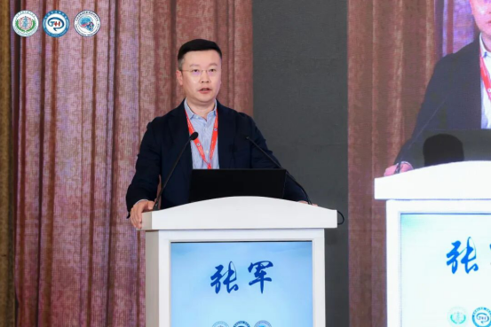
Professor Zhang Jun focused on the surgical management of giant vestibular schwannomas, analyzing the technical challenges associated with these complex tumors. He discussed endoscope-assisted microsurgical strategies and adjuvant radiotherapy for postoperative residual tumors, placing particular emphasis on facial nerve preservation. His presentation highlighted the importance of achieving maximal safe tumor resection while minimizing the risk of facial paralysis and other serious postoperative neurological complications.
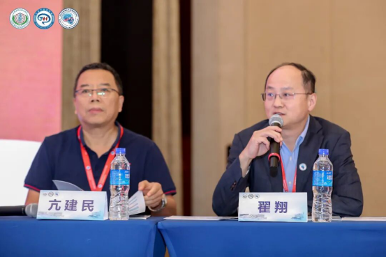
The scientific presentation session was co-chaired by Professors Zhang Xiaohua, Zhai Xiang, and Kang Jianmin.
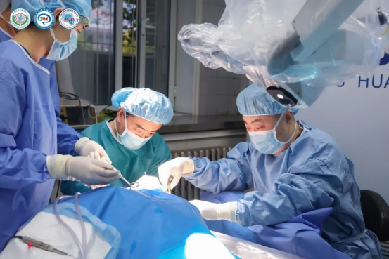
Professor Tong Xiaoguang and Director Shi Minggang concluded the session with a presentation entitled “The Kawase Approach and Fukushima Bypass.” Through hands-on anatomical demonstrations illustrating the application of the Fukushima bypass in skull base surgery, they systematically reviewed the key technical considerations and innovative value of this approach in complex skull base reconstruction. They provided a detailed overview of the clinical applications of Fukushima bypass techniques in the surgical management of large skull base tumors and complex intracranial aneurysms. In cases involving major cerebral vessels, vascular bypass and reconstruction can help maintain adequate cerebral perfusion, thereby improving the extent of tumor resection and enhancing the safety of complex skull base procedures.
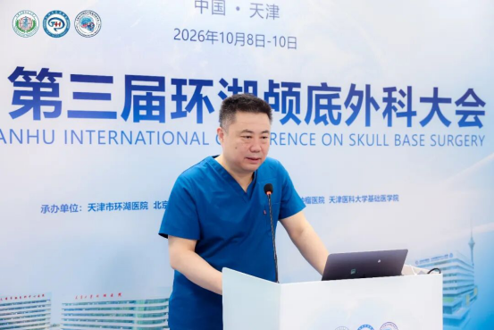
As the conference drew to a close, Professor Tong Xiaoguang and Professor Juan Fernandez-Miranda delivered a concise review of the day's scientific sessions. They emphasized that the future development of neurosurgery requires a stronger commitment to the integrated framework of “Anatomy–Imaging–Surgery,” fostering deeper integration of cutting-edge technologies with clinical practice.
The conference was not only a distinguished platform for academic exchange but also a milestone in the convergence of innovative ideas and surgical advancements. The participating experts shared the view that, driven by the complementary forces of precision medicine and intelligent technologies, skull base surgery is entering a new era characterized by greater safety, enhanced efficiency, and more patient-centered care.